Admission
The patient was admitted into hospital at 8:00am on Monday 13 September 2010. The anaesthetic consent form (see appendix) was explained to the owner and she was asked if Tessa had eaten since 8:00pm the previous evening and if she had been prescribed any medication recently to which the owner circled no for both questions however noted that Tessa had one dose of Metacam on Friday 10 September 2010. The form was then signed and dated. It was also noted on the form that if there are any metastasis found while the patient is under anaesthesia then please euthanize. This comment was also signed and dated. Tessa was then weighed and her weight was recorded onto her electronic file and the anaesthetic consent form. It was important to weigh Tessa to ensure that all the medication calculations were accurate.
Pre-Operative Care
 |
| Image 12 |
subcutaneously (see appendix). She was then taken for a walk so that she could relieve herself and vomit if need be. After returning from her walk, an intravenous catheter was placed into the right cephalic vein and at 9:00am, 1.8mLs of Cephazolin Sodium[3] (see appendix) was administered slowly intravenously through the catheter. It was noted that the Cephazolin Sodium[3] would need to be repeated 8 hourly until it was stopped and the last dose given at 12:00pm on Tuesday 14 September 2010. The patient was clipped 25cm cranial to the sacrum and 9cm lateral to the spine. The initial preparation was done using cotton balls with Chlorhexidine Gluconate[4] and water ensuring that the entire clip sight was clean and any hair or dirt was removed. Artificial tears were placed in the patient’s eye before she was taken through to theatre to prevent drying of the eye. Once the patient was taken through to theatre, the
 |
| Image 13 |
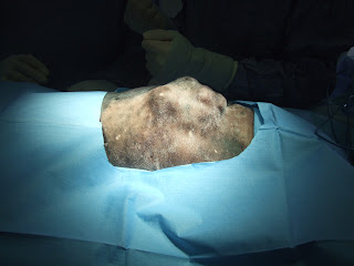
final sterile preparation was completed using sterile swabs, Chlorhexidine Gluconate[4] and sodium for irrigation[5] in a sterile bowl. A concentric circle motion technique was used to ensure that any remaining dirt was moved away from the potential surgical incision site. Once the preparation was complete, sterile drapes were placed over the patient (see image 12 and 13) while maintaining an aseptic technique and ensuring that the drapes did not touch any of the unsterile equipment. The veterinarian and assistant nurse then scrubbed into surgery and gowned and gloved using the aseptic technique.
During the Surgical Procedure
During the surgical procedure, it was important that the patient’s anaesthetic was constantly monitored. One of the nurses monitored the anaesthetic of the patient and recorded all the findings every 5 minutes on the anaesthetic record sheet (see appendix). Intermittent positive pressure ventilation was provided to the patient during part of the surgical procedure when the thoracic cavity was open and negative pressure was not able to be achieved. The nurse that was scrubbed into the surgery directly assisted the veterinarian with blotting blood using a sterile swab, holding retractors, flushing the wound, handing over instruments and cutting the sutures. Another nurse was present to open any of the kits or extra instruments that the veterinarian needed. When these were opened, an aseptic technique was used to pass the product or instrument to the veterinarian.
Post-Operative Care
 |
| Image 14 |
Once the surgical procedure was complete and the patient was waiting to be extubated (see image 14), a light sterile thoracic bandage was placed around the patient’s thorax. It was made of soffban and vetrap and small cuts were placed around the bandage to prevent any possible pressure build up and to allow more comfort for the patient. The patient woke up at 1:00pm and was very vocal. At this time, 0.08mLs of Acepromazine[6] (see appendix) was administered intravenously to help calm her down. It was noted to administer this as required and a repeat administration was given at 3:00am the following day (Tuesday 14 September 2010). At this time, she was also administered with a one off dose of 0.66mLs of Carprofen[7] (see appendix) subcutaneously for pain relief. During the surgical procedure, a closed suction drain was placed in order to remove any fluid that may build up in the thoracic cavity following the surgery. It was reported that it needed to be checked every 4 hours. At 1:00pm, 10mLs was removed through the drain, at 5:00pm, 20mLs was removed, at 6:00pm, 5mLs was removed and at 8:00am the following day (Tuesday 14 September 2010), 40mLs was removed and the drain was removed completely at 11:00am the same day. A chest tube was also placed during the procedure and it needed to be checked every 4 hours also. At 12:00pm, negative pressure was still present within the thoracic cavity, at 1:00pm, negative pressure remained, at 3:00pm 1mL of fluid was removed and at 6:00pm pressure was negative also. The tube was checked again at 8:00am the following day (Tuesday 14 September 2010) and the pressure remained negative and the tube was removed at 11:00am. Once the patient was fully recovered, her chest drain and chest tube were checked regularly and her medications were administered as required according to the daily record sheets (see appendix). She was fed soft food, usually chicken and rice twice daily and was walked 4-6 times daily for 5 minutes just enough to allow her to relieve herself. This general care was maintained up until she was discharged at 5:00pm on Friday 17 September 2010.
No comments:
Post a Comment